
Ultrasound-Guided Regional Anaesthesia: Why Visualization Matters
Founder & Clinical Director, Ultrascan Technologies
March 15, 2026 - 8 min read
Regional anaesthesia has been transformed by ultrasound guidance. Where landmark-based techniques relied on knowledge of surface anatomy and the subjective feel of tissue planes, ultrasound guidance makes the needle, the nerve, and the surrounding structures visible in real time. The clinical outcomes are better, the complication rates are lower, and the required local anaesthetic volumes are reduced. For any clinician performing peripheral nerve blocks, the question is no longer whether to use ultrasound - it is which device best fits the workflow.
The Evidence Base for Ultrasound-Guided Regional Anaesthesia
The European Society of Regional Anaesthesia and Pain Therapy (ESRA) guidelines strongly endorse ultrasound guidance for peripheral nerve blocks. Their position, supported by multiple meta-analyses, is that real-time ultrasound guidance improves block success rates, reduces onset time, and decreases the risk of vascular puncture, intraneuronal injection, and local anaesthetic systemic toxicity (LAST).
A landmark meta-analysis published in Anaesthesia comparing ultrasound versus nerve stimulation for peripheral nerve blocks found that ultrasound guidance reduced procedure time by a median of 38%, improved block success rates by a relative 11%, and significantly reduced the risk of vascular puncture. For upper limb blocks, the benefit was most pronounced in the brachial plexus approaches.
For reference, the ESRA position statement on ultrasound-guided regional anaesthesia guidelines is publicly available and provides a comprehensive evidence synthesis.
Key Nerve Blocks and How Ultrasound Changes Them
Femoral Nerve Block
The femoral nerve lies lateral to the femoral artery in the femoral triangle, beneath the fascia iliaca. On ultrasound, the nerve appears as a hyperechoic (bright) oval or triangular structure, typically 1-2 cm lateral to the pulsating femoral artery. Without ultrasound, landmark technique deposits local anaesthetic medially near the artery or requires a "click" through the fascia - a technique with significant interoperator variability.
With ultrasound, you can confirm needle tip position adjacent to the nerve before injection, watch the spread of local anaesthetic (which should appear as anechoic fluid encircling the nerve), and immediately identify inadvertent intravascular needle placement by the lack of tissue expansion and the presence of vessel compression artefact. The typical dose for a femoral nerve block decreases from 20-30 mL with landmark technique to 10-15 mL under ultrasound guidance, reducing the risk of LAST without sacrificing block quality.
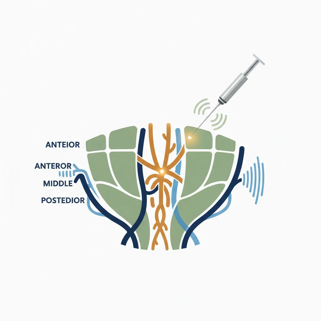
Brachial Plexus: Interscalene Approach
The interscalene block targets the roots of the brachial plexus between the anterior and middle scalene muscles at the C5-C6 level. The roots appear as hypoechoic oval structures stacked between the scalene muscles in short-axis view. The critical adjacent structures - the internal jugular vein, carotid artery, and phrenic nerve - are all visible and avoidable under real-time guidance.
Phrenic nerve palsy (causing ipsilateral hemidiaphragm paralysis) occurs in virtually all interscalene blocks regardless of technique, but the incidence of clinically significant respiratory compromise is reduced with lower local anaesthetic volumes guided by ultrasound confirmation of spread.
Brachial Plexus: Supraclavicular Approach
The supraclavicular approach targets the trunks and divisions of the brachial plexus as they cluster above the first rib and lateral to the subclavian artery. This is the "spinal of the arm" - excellent coverage for forearm and hand surgery. Without ultrasound, pneumothorax risk from blind supraclavicular techniques was significant enough that many anaesthetists avoided the approach entirely. With ultrasound, the rib, pleural line, and subclavian artery are all visible, making the approach safer. Studies have shown pneumothorax rates below 0.5% with ultrasound guidance versus 1-6% with landmark technique.
Sciatic Nerve Block
The sciatic nerve is the largest peripheral nerve in the body and can be approached at multiple levels - subgluteal, mid-thigh, or popliteal. At the popliteal level, the nerve appears as a large hyperechoic structure just before it bifurcates into the tibial and common peroneal nerves. Ultrasound allows confirmation that both components are encircled by local anaesthetic before injection is complete.
Transversus Abdominis Plane (TAP) Block
The TAP block deposits local anaesthetic in the fascial plane between the internal oblique and transversus abdominis muscles, providing anterior abdominal wall analgesia for laparotomy, hernia repair, and caesarean section. This plane is not palpable and cannot be reliably accessed without imaging. Ultrasound makes TAP a reliable, reproducible technique - you watch the layers separate as local anaesthetic spreads in the correct plane, confirming successful injection before the patient leaves the anaesthetic room.
Local Anaesthetic Volume Reduction
One of the most clinically important benefits of ultrasound-guided regional anaesthesia is the reduction in required local anaesthetic volumes. When you can confirm that the needle tip is adjacent to the target nerve and watch injectate spread, you need significantly less volume to achieve circumferential nerve coverage compared to landmark techniques where approximate position necessitates generous volumes.
This matters because total local anaesthetic dose is the primary determinant of LAST risk. Reducing volumes by 30-50% while achieving equivalent or superior block quality directly reduces the risk of this potentially life-threatening complication.
Handheld Ultrasound for Regional Anaesthesia
A wireless handheld probe is entirely appropriate for most standard peripheral nerve blocks. The linear probe provides the high-frequency, high-resolution imaging needed for superficial nerve identification. The key requirements are adequate image depth (typically 3-5 cm for most brachial plexus approaches, up to 8-10 cm for sciatic nerve at the gluteal level), a linear array for parallel needle tracking, and the ability to hold a stable image while managing the needle simultaneously.
The Ultrascan US-PL combines a phased array element for cardiac and deeper structures with a linear element suited to regional anaesthesia and vascular access. For an anaesthetist who also performs bedside cardiac assessment and vascular access, this dual capability makes it the most versatile single-probe option in the Ultrascan range.
For training in ultrasound-guided regional anaesthesia techniques, explore the Ultrascan education resources, or visit our specialties pages for anaesthesia-specific guidance on device selection and application.
Visual Summary
Key concepts from this article at a glance.

Related Products
Related Articles
POCUS in Anaesthesia: Vascular, Gastric, Lung and Cardiac
A focused review of point-of-care ultrasound applications in anaesthesia practice.
Ultrasound-Guided Vascular Access: Where It Adds Value
Evidence-based review of ultrasound guidance for peripheral and central vascular access.

Practical Guide to FAST and eFAST with Handheld Ultrasound
Step-by-step guide to performing FAST and extended FAST assessments using portable ultrasound.