POCUS in Anaesthesia: Vascular, Gastric, Lung and Cardiac
Founder & Clinical Director, Ultrascan Technologies
7 March 2026 - 11 min read
Dr. Yahya Docrat, MBChB
Founder and Clinical Director at Ultrascan Technologies. Anaesthesiologist and POCUS educator with clinical practice across South Africa.
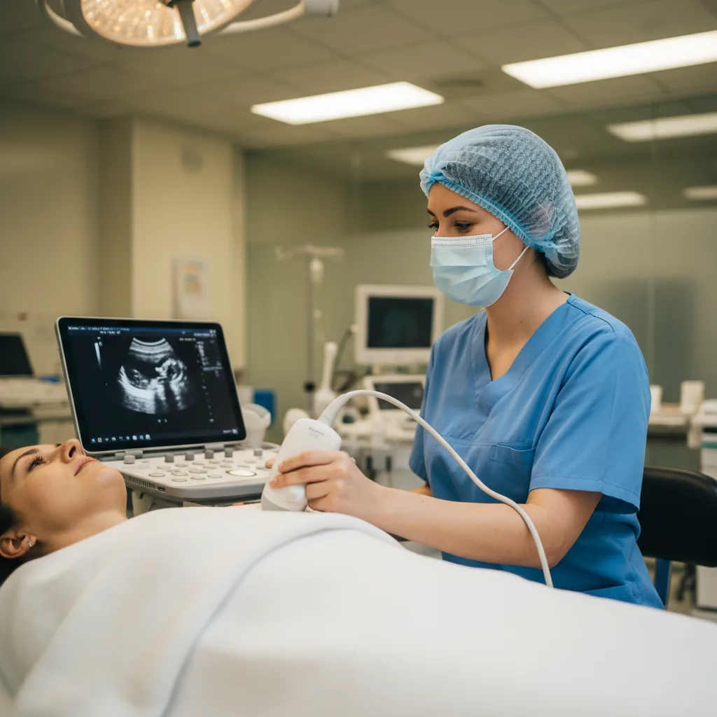
Point-of-care ultrasound has found a natural home in anaesthesia practice. The anaesthetist is already at the bedside, already performing procedures, and already making time-sensitive decisions about airway, circulation, and fluid management. Adding a handheld probe to this workflow does not require a new clinical paradigm - it requires understanding which questions ultrasound can answer in the peri-operative context and integrating those answers into decisions that were previously made on less information.
This article covers the four main POCUS applications in anaesthesia: pre-operative cardiac assessment, gastric ultrasound for aspiration risk, lung ultrasound, and procedural guidance including vascular access and regional anaesthesia.
Pre-operative Cardiac Assessment
Many patients presenting for surgery have not had formal echocardiography, and their cardiac status is inferred from history, examination, ECG, and functional capacity assessment. A focused cardiac assessment with a handheld probe in the pre-operative room takes five to seven minutes and can identify:
- Severely reduced LV function not previously documented
- Significant pericardial effusion
- Dilated right ventricle suggesting pulmonary hypertension or chronic cor pulmonale
- Gross valvular abnormality (restricted aortic valve, flail mitral leaflet)
- Left ventricular hypertrophy suggesting long-standing hypertension
Any of these findings may alter the anaesthetic plan - fluid strategy, choice of anaesthetic agents, monitoring requirements, or the decision to delay the procedure for formal cardiac evaluation. The British Journal of Anaesthesia has published guidelines on POCUS in anaesthesia, available through the BJA online platform, which endorse focused cardiac assessment as a valuable pre-operative tool when formal echocardiography has not been performed.
The Ultrascan US-PL phased array is the appropriate probe for pre-operative cardiac assessment. Its phased array footprint fits between rib spaces without the acoustic shadowing that linear probes produce.
Gastric Ultrasound for Aspiration Risk
Pulmonary aspiration remains one of the most feared complications in anaesthesia. Standard fasting guidelines (six hours for solids, two hours for clear fluids) are designed to ensure gastric emptying before induction, but they are not reliable in all patients. Delayed gastric emptying is common in diabetic patients, patients with opioid use, trauma patients who have eaten recently, and obstetric patients.
Gastric ultrasound provides a direct assessment of gastric content and volume at the time of anaesthetic induction. The technique images the antrum of the stomach in the right lateral decubitus position using a convex probe. An empty stomach (grade 0) shows a small, collapsed antrum. A stomach containing clear fluid (grade 1) shows fluid in one position only. A stomach with solid or mixed content (grade 2) shows content in both supine and lateral positions.
Gastric antral cross-sectional area (CSA) measured in the right lateral decubitus position correlates with gastric volume. A validated formula (Perlas equation) uses the CSA measurement to estimate volume, with a CSA above 340 mm2 generally indicating a gastric volume above 1.5 mL/kg (the threshold associated with aspiration risk).
A systematic review published in Anesthesiology on gastric ultrasound for aspiration risk assessment found that the technique was feasible in over 95% of patients, with strong correlation between ultrasound-estimated and actual gastric volumes. The review supported gastric ultrasound as a reliable pre-induction assessment tool in high-risk patients.
For the anaesthetist, gastric ultrasound takes under three minutes in most patients and directly informs the decision between standard induction and rapid sequence induction (RSI).
Lung Ultrasound in the Peri-operative Setting
Lung ultrasound has been validated in multiple studies as superior to chest X-ray for detecting pleural effusion, pulmonary consolidation, and pneumothorax. In the anaesthetic context, three applications are most relevant:
Pre-operative Lung Assessment
In patients with respiratory symptoms or signs, a pre-operative lung ultrasound can identify pleural effusions, consolidation, or interstitial syndrome (B-lines indicating pulmonary oedema) that may alter the anaesthetic plan. A large pleural effusion identified pre-operatively allows planning for drainage before surgery or adjustment of ventilation strategy.
Post-operative Atelectasis
Atelectasis is the most common post-operative pulmonary complication and appears on lung ultrasound as subpleural consolidation with air bronchograms in the dependent lung zones. Identifying atelectasis by ultrasound allows targeted physiotherapy, positioning changes, and recruitment manoeuvres rather than empirical management of undifferentiated hypoxaemia.
Intra-operative Monitoring
In prolonged procedures or those with risk of pneumothorax (thoracic cases, subclavian access, high-pressure ventilation), lung ultrasound provides a rapid check for iatrogenic pneumothorax that can be performed without interrupting surgery or exposing the patient to radiation.
Vascular Access and Regional Anaesthesia
Vascular access guidance and regional anaesthesia are discussed in depth in separate articles on this site. In the anaesthetic context, the key point is workflow integration: the probe used for pre-operative cardiac and gastric assessment can be the same probe used for vascular access immediately before induction and for nerve block guidance in the anaesthetic room. A single handheld device covers multiple applications in sequence without changing setup.
For procedural guidance specifically, the linear element of the US-PL provides the high-frequency imaging needed for nerve and vessel visualisation. The phased array element handles cardiac assessment in the same session.
How Handheld Devices Fit Anaesthesia Workflow
The anaesthetic workflow has specific constraints that influence which ultrasound platform is practical. Setup time matters in a busy list. Probe hygiene between patients matters in an environment with high sterility requirements. Portability matters when you are moving between the anaesthetic room, the theatre, and the recovery bay.
A wireless handheld probe addresses these constraints. There are no cables to manage around an anaesthetic machine. The probe can be sleeved for sterile use during procedures and cleaned between patients with approved surface disinfectants. The app-based workflow on a dedicated tablet or secured phone means images can be saved to the patient record without a separate workstation.
For anaesthesia departments considering the transition from cart-based or shared machines to individual handheld probes, the Ultrascan specialties pages provide application-specific guidance, and the education resources include training pathways for peri-operative POCUS skills.
POCUS in anaesthesia is not a replacement for comprehensive echocardiography or formal radiological reporting. It is a focused, time-critical assessment tool that answers specific clinical questions at the moment they arise. Used within its scope, it directly improves peri-operative decision-making and patient safety.
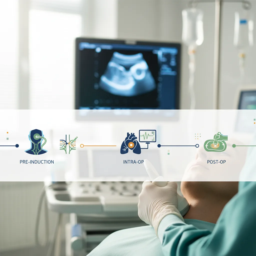
Visual Summary
Key concepts from this article at a glance.

Related Products
Related Articles

Point-of-Care Ultrasound in South African Practice
How POCUS is transforming clinical practice across South Africa, from rural clinics to urban emergency departments.

Practical Guide to FAST and eFAST with Handheld Ultrasound
Step-by-step guide to performing FAST and extended FAST assessments using portable ultrasound.

Ultrasound-Guided Regional Anaesthesia: Why Visualization Matters
The case for ultrasound guidance in nerve blocks and regional anaesthesia techniques.