Ultrasound-Guided Vascular Access
Overview
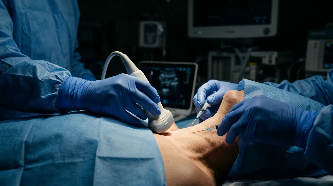
Real-time ultrasound guidance for vascular access has become the standard of care for central venous catheterisation and is increasingly used for peripheral access in difficult patients. This guide covers technique for internal jugular, subclavian, femoral, and peripheral access under ultrasound.
Founder & Clinical Director, Ultrascan Technologies
1 November 2025 - 7 min read
Dr. Yahya Docrat, MBChB
Founder and Clinical Director at Ultrascan Technologies. Anaesthesiologist and POCUS educator with clinical practice across South Africa.
Key Clinical Points
Two approaches: real-time in-plane (long axis) and out-of-plane (short axis, most common)
The needle tip must be visualised at all times to prevent posterior wall puncture
Compress the vein to confirm arterial versus venous anatomy before cannulation
Dynamic needle tip visualisation improves first-pass success rates significantly
Linear probe provides highest resolution for peripheral access in difficult veins
Confirm wire position in the vessel before dilation for central access
Related Specialties
Anaesthesiology
Pre-operative assessment, regional nerve block guidance, and airway ultrasound for safe anaesthetic delivery.
Emergency Medicine
Rapid bedside assessment for trauma, haemodynamic instability, and time-critical diagnoses in the emergency department.
Vascular
Real-time vascular imaging for access guidance, DVT diagnosis, and peripheral arterial assessment in clinical and procedural settings.
Critical Care / ICU
Comprehensive critical care ultrasound for mechanically ventilated patients, haemodynamic instability, and procedural guidance in the ICU.
Ready to put this into practice?
Book a free hands-on POCUS demonstration with Dr. Docrat at your practice.
Book a Free Demo