Bedside Cardiac Ultrasound: What Handheld Can and Cannot Tell You
Founder & Clinical Director, Ultrascan Technologies
13 March 2026 - 9 min read
Dr. Yahya Docrat, MBChB
Founder and Clinical Director at Ultrascan Technologies. Anaesthesiologist and POCUS educator with clinical practice across South Africa.
Focused cardiac ultrasound (FoCUS) has become one of the most requested point-of-care skills in emergency medicine, critical care, and internal medicine. The ability to assess gross left ventricular (LV) function, detect a pericardial effusion, or evaluate volume status at the bedside has genuine clinical impact. But handheld and portable ultrasound devices have real limitations compared to formal transthoracic echocardiography (TTE), and understanding those limits is as important as knowing what they can do.
This article covers what focused cardiac ultrasound with a handheld device can reliably answer, where it falls short, and how to incorporate FoCUS into your clinical reasoning without overextending its diagnostic reach.
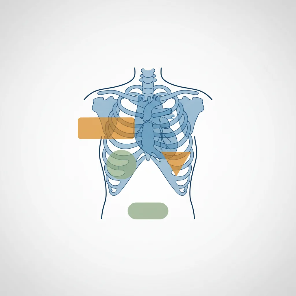
The Four Standard Cardiac Views
Parasternal Long-Axis (PLAX)
Probe position: 3rd to 4th intercostal space, left sternal edge, indicator pointing to the patient's right shoulder. This view shows the left ventricle, left atrium, mitral valve, aortic valve, and the proximal aorta in a single long-axis plane. PLAX is the starting point for most FoCUS assessments. LV chamber size, gross wall motion, and the presence of pericardial effusion are assessed here.
Parasternal Short-Axis (PSAX)
Rotate 90 degrees clockwise from PLAX. The left ventricle appears as a circular structure in cross-section. At the papillary muscle level, you can assess regional wall motion abnormalities in multiple coronary artery territories simultaneously. A D-shaped septum (the septum bowing into the left ventricle) indicates right ventricular pressure or volume overload.
Apical Four-Chamber (A4C)
Probe position: cardiac apex (usually the point of maximal impulse), indicator pointing to the patient's left. This view shows all four chambers simultaneously and is the best single view for assessing relative chamber sizes and global LV function. RV dilation (RV to LV ratio greater than 0.6 in this view) raises concern for right heart strain. Global LV systolic function is often most intuitive to assess in this view.
Subxiphoid (Subcostal)
As described in the FAST protocol, this view provides an alternative window to the cardiac chambers and pericardium. In patients with poor parasternal windows (obese, barrel-chested, hyperinflated lungs), the subcostal view may be the only feasible approach. IVC assessment is performed from the subxiphoid approach by angling slightly to the patient's right.
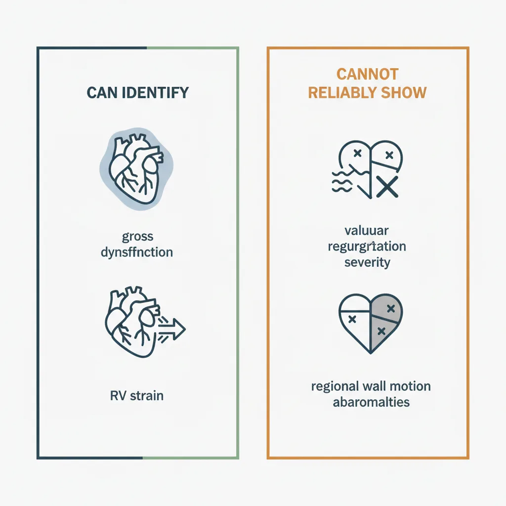
What FoCUS Can Reliably Tell You
Global LV Function
Visual estimation of LV ejection fraction (EF) by an experienced operator correlates well with formal echocardiographic measurement. The standard classification used in FoCUS is a three-tier system: normal (roughly above 50%), moderately reduced (roughly 30-50%), and severely reduced (below 30%). This categorisation is adequate for most acute clinical decisions.
A study published in JACC Cardiovascular Imaging demonstrated that handheld cardiac ultrasound performed by trained operators showed good agreement with formal TTE for detecting reduced LV function, with sensitivity above 85% for detecting EF below 40%.
Pericardial Effusion
FoCUS is highly sensitive for detecting pericardial effusion. Free fluid appears as an anechoic space around the heart. Location and circumferential extent help estimate severity. Early tamponade physiology - right atrial systolic collapse and right ventricular diastolic collapse - can be identified with experience. In the context of shock, a large effusion with right-sided chamber collapse is a diagnosis that changes immediate management.
Right Ventricular Dilation
In the setting of acute dyspnoea or unexplained haemodynamic instability, a dilated RV with septal flattening (D-sign on PSAX) raises concern for massive pulmonary embolism, severe pulmonary hypertension, or RV infarct. This finding in the appropriate clinical context should prompt urgent investigation and management.
IVC Assessment for Volume Status
The inferior vena cava (IVC) diameter and its respiratory variation provide a surrogate for right atrial pressure. A small, collapsing IVC (diameter below 15 mm with greater than 50% respiratory collapse) suggests low right atrial pressure and volume responsiveness. A large, non-collapsing IVC suggests elevated right atrial pressure. This assessment is most reliable in spontaneously breathing patients and loses validity in mechanically ventilated patients.
What Handheld FoCUS Cannot Tell You
This section is as important as the capabilities above. Overextending the diagnostic scope of handheld cardiac ultrasound is a patient safety issue.
The American Society of Echocardiography (ASE) guidelines on focused cardiac ultrasound, available at the ASE guidelines portal, clearly delineate the boundary between FoCUS and comprehensive echocardiography.
Valvular Disease
Handheld devices used for FoCUS typically do not include colour Doppler or spectral Doppler in a form that allows reliable valvular assessment. You may detect gross aortic stenosis from a heavily calcified, restricted valve, or severe mitral regurgitation from a flail leaflet, but quantification of valvular lesions requires spectral Doppler measurements that FoCUS is not designed to provide. Any suspicion of significant valvular disease on FoCUS should prompt formal echocardiographic evaluation.
Diastolic Function
Diastolic assessment requires tissue Doppler imaging and transmitral flow velocity measurements. These are beyond the scope of standard handheld FoCUS. Do not attempt to characterise diastolic function from handheld images without the necessary Doppler tools and training.
Strain Imaging and Advanced Measurements
Global longitudinal strain (GLS) and other advanced myocardial mechanics assessments require dedicated echocardiography software. Handheld FoCUS is not a platform for these measurements, regardless of device.
Congenital Abnormalities
Complex congenital cardiac anatomy requires systematic, comprehensive imaging with multiple views and measurements. FoCUS will not reliably detect or characterise congenital abnormalities beyond gross structural changes.
Integrating FoCUS Into Clinical Practice
FoCUS is designed to answer specific binary questions in a clinical context where the answer changes immediate management. The framework: is there significant LV dysfunction? Is there a pericardial effusion with tamponade physiology? Is there RV dilation suggesting acute cor pulmonale? Is this patient volume-depleted?
When the answer to any of these questions is "yes" or "I cannot rule it out," the appropriate response is to escalate - either to immediate management for clear findings, or to formal echocardiography for equivocal ones.
For phased array cardiac imaging from a handheld device, the Ultrascan US-PL is the recommended choice. Its phased array element is designed for cardiac imaging windows, and the linear element provides the additional vascular access capability useful in critical care.
For training resources on FoCUS and other POCUS applications, visit the Ultrascan education page. For clinical questions about suitability for your practice, the FAQ page covers common queries about handheld cardiac imaging.
Visual Summary
Key concepts from this article at a glance.