How to Choose the Right Wireless Ultrasound Probe
Founder & Clinical Director, Ultrascan Technologies
March 3, 2026 - 10 min read
Choosing a wireless ultrasound probe is a more consequential decision than it might first appear. The probe you select will shape which patients you can assess, which clinical questions you can answer, and how effectively you can integrate ultrasound into your daily workflow. Get it right and POCUS becomes an effortless extension of clinical examination. Get it wrong and you will find yourself with a device that does not cover your core use cases, or one that produces frustrating images in the body regions you scan most.
This guide walks through probe selection systematically: probe types, clinical applications by specialty, key technical specifications, and practical considerations for South African practitioners.
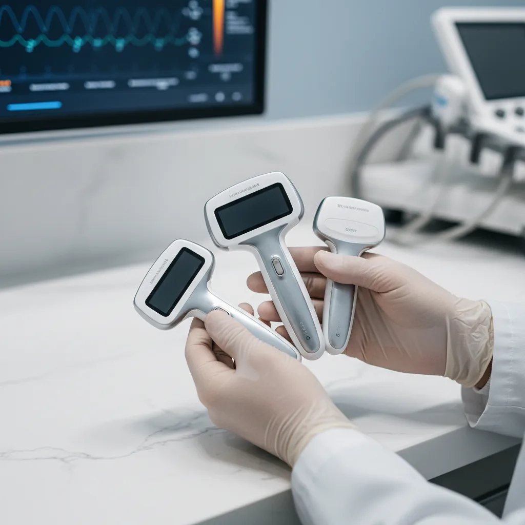
Understanding Probe Types
Convex (Curvilinear) Probes
The convex probe is the most versatile general-purpose probe in clinical POCUS. Its curved array produces a wide sector image with a broad field of view at depth, making it ideal for abdominal organs, obstetric assessment, and deep thoracic structures. Typical frequencies range from 2 to 5 MHz, favouring penetration over resolution. If you can only have one probe and your work involves general medicine, emergency, obstetrics, or rural practice, the convex probe covers the widest range of clinical scenarios.
The Ultrascan US-CE is a wireless convex probe particularly suited to abdominal and obstetric applications. It connects directly to a smartphone app and requires no base station or cart.
Linear Probes
Linear probes produce a rectangular image with high resolution at shallower depths, typically 1 to 5 cm. They operate at higher frequencies, usually 7 to 15 MHz, which yields excellent near-field detail. Linear probes are the correct choice for vascular access (peripheral and central veins), superficial soft tissue, nerve identification for regional blocks, musculoskeletal assessment, and thyroid or breast evaluation.
If your primary POCUS use cases are procedural guidance (line placement, nerve blocks, joint aspirations) or assessment of superficial structures, a linear probe is essential. It cannot substitute for convex at depth, but no other probe can match it for superficial work.
Phased Array (Cardiac) Probes
The phased array probe has a small footprint that fits between ribs, making it the probe of choice for cardiac imaging. It produces a sector-shaped image with a wide far field from a narrow contact surface. Frequencies are typically 1 to 5 MHz. Phased array probes are used for focused cardiac assessment (parasternal, apical, and subcostal views), lung sliding assessment from the chest wall, and diaphragm visualisation.
Emergency physicians, intensivists, and anaesthesiologists who need cardiac assessment will require either a dedicated phased array probe or a dual-probe device that includes phased array capability. General practitioners performing abdominal and obstetric scanning do not routinely need it.
Dual-Head and Multi-Frequency Probes
Several wireless probe platforms offer a dual-frequency design in a single device, typically combining a convex and linear array in one probe. This significantly broadens clinical versatility without requiring the clinician to carry multiple probes. The trade-off is slightly more physical bulk compared to a single-element device, and some dual-head designs represent compromises in image quality relative to dedicated single-use probes.
The Ultrascan US-CL dual-head probe addresses this directly, offering convex and linear functionality in one wireless device. For clinicians who need both abdominal and vascular access capability, this represents compelling value. The US-CL Pro extends this with enhanced image processing for higher-demand clinical environments.
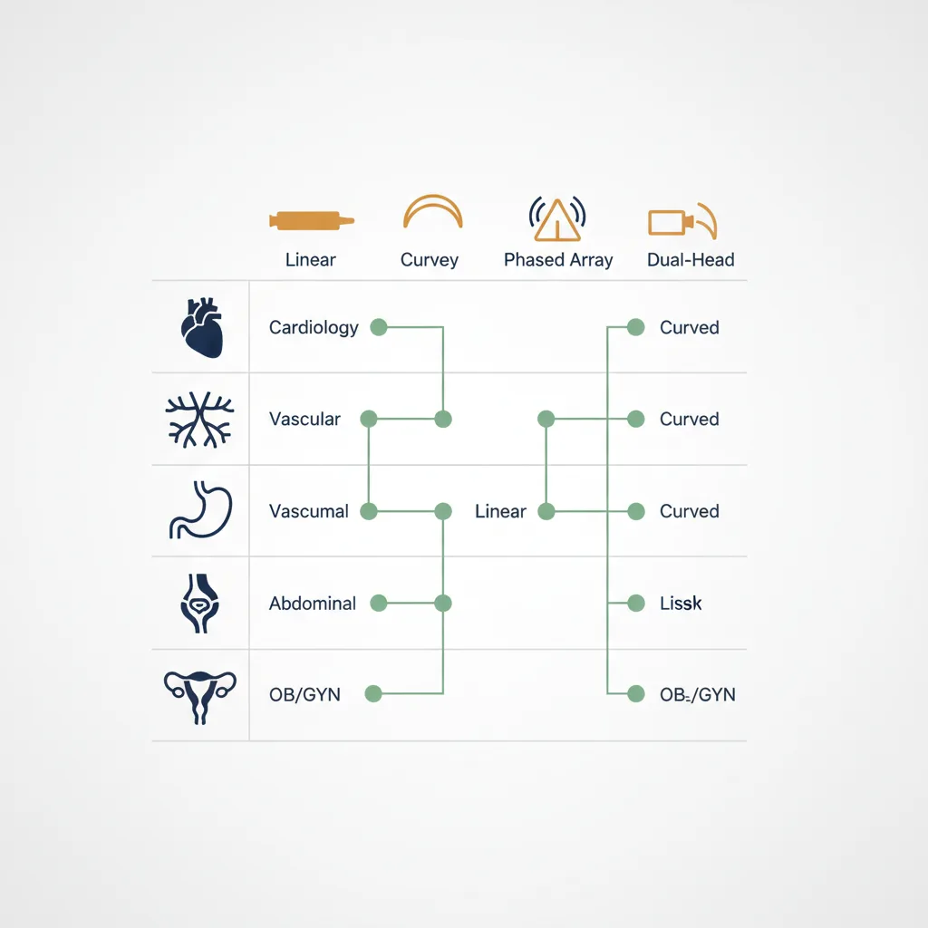
Matching Probe to Specialty
- Emergency medicine: Dual convex-linear probe as minimum; phased array preferred for full cardiac and lung protocol capability
- General practice and family medicine: Convex probe covers the majority of use cases (abdomen, obstetrics, DVT screening)
- Anaesthesia: Linear probe essential for vascular access and nerve blocks; phased array useful if performing peri-operative cardiac assessment
- Intensive care: All three probe types useful; dual-head devices reduce complexity on busy rounds
- Obstetrics and gynaecology: Convex probe for abdominal; endovaginal transducer for first trimester and pelvic assessment
- Musculoskeletal and sports medicine: Linear probe is primary tool
- Rural and district hospital medicine: Dual convex-linear probe offers maximum versatility for unknown case mix
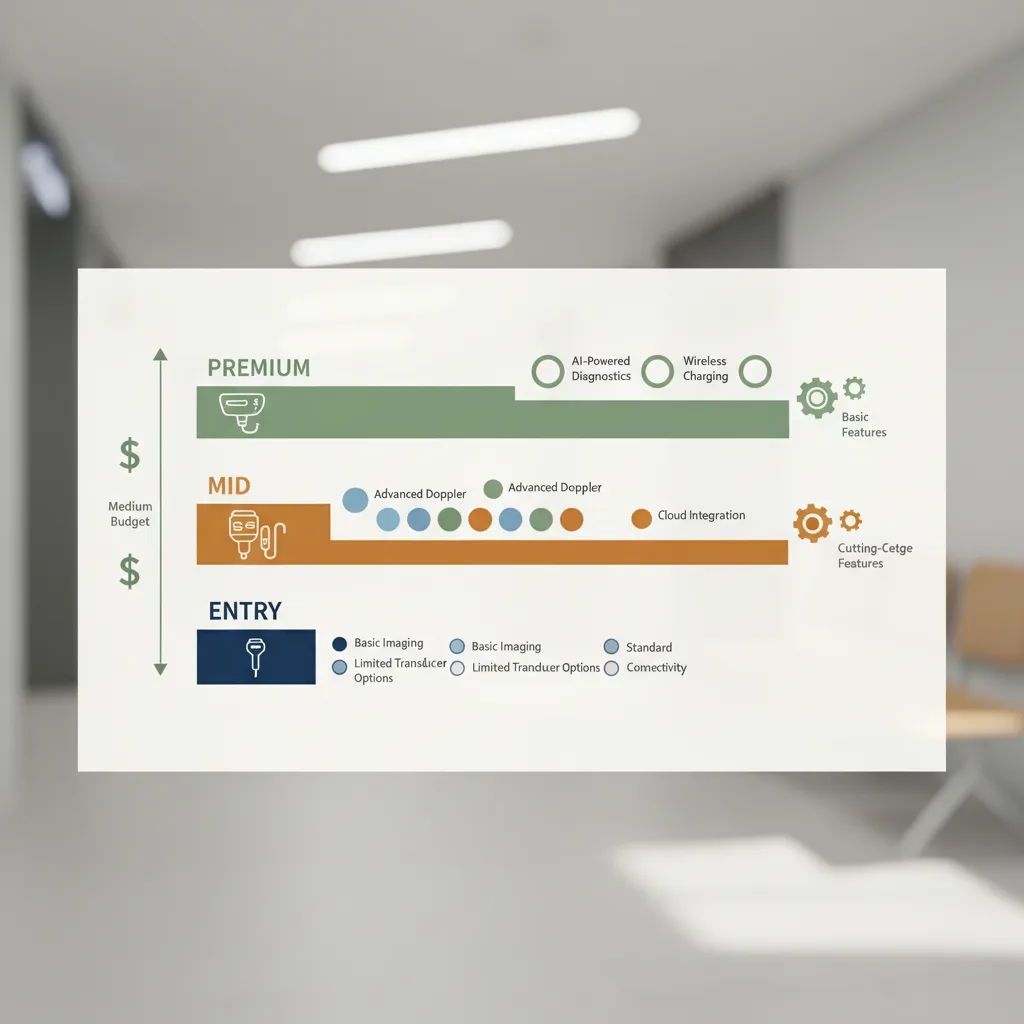
Key Technical Specifications to Evaluate
Frequency Range
Broadband probes that span a wide frequency range give you more flexibility. A probe rated 2 to 8 MHz can be tuned for deep penetration in obese patients or switched to higher frequency for sharper superficial detail. Single-frequency probes are more limited. Look for broadband specifications rather than single nominal frequencies.
Imaging Depth
Most wireless probes claim maximum depths of 15 to 25 cm for convex arrays. In clinical practice, obese patients require adequate penetration at depth with acceptable resolution. Review independent image quality assessments rather than relying solely on manufacturer specifications.
Battery Life
Wireless probe battery life is often cited between 1 and 3 hours per charge. For clinicians doing short, targeted scans across a morning clinic, 1 hour may be sufficient. For emergency physicians or intensivists doing extended assessment sessions, 2 to 3 hours of continuous use is meaningfully better. Assess this against your expected usage pattern.
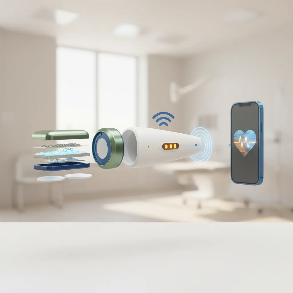
Connectivity and App Compatibility
Wireless probes connect via WiFi direct or Bluetooth to smartphone apps. Assess whether the app works on the devices your team already carries (iOS vs Android), whether it stores images locally, and whether it integrates with your practice management system. Some premium applications include AI-assisted image guidance, which can be valuable for clinicians in the early stages of their POCUS learning curve.
Subscription Fees: The Hidden Cost
Many wireless probe manufacturers charge annual subscription fees for full app functionality, cloud storage, or software features. These fees range from approximately R8,000 to R30,000 per year depending on the platform. Over five years, this can add R40,000 to R150,000 to the total cost of ownership on top of the purchase price.
All Ultrascan devices carry no subscription fees. The probe price is the total price. For a more detailed breakdown, see our US-CL Pro product page or the US-PL phased-linear probe.
External Evidence on Probe Selection
The World Federation for Ultrasound in Medicine and Biology (WFUMB) has published a position statement on portable ultrasound, noting that probe selection should be guided primarily by the clinical context and application rather than by brand or platform. A review of wireless probe performance published in the Journal of Ultrasound in Medicine found that diagnostic accuracy was closely linked to probe-frequency matching for the target anatomy, reinforcing the importance of correct probe selection over raw image quality metrics. The WFUMB portable ultrasound position statement provides a useful framework for practitioners making this decision.
Making the Decision
For most South African practitioners new to POCUS, a dual convex-linear probe represents the best starting point. It covers the broadest range of clinical applications without requiring multiple purchases, and eliminates the need to select between probes mid-assessment. As your POCUS practice develops and specific gaps emerge, a second specialised probe can be added.
Explore the full specialty application guide to see specific probe recommendations by clinical area, or contact the Ultrascan team to discuss which device matches your clinical environment.
Visual Summary
Key concepts from this article at a glance.
Related Articles
Ultrascan vs Clarius: Which Wireless Probe is Right for You?
A detailed comparison of Ultrascan and Clarius wireless ultrasound probes for SA practitioners.

The True Cost of POCUS: A ZAR Pricing Guide for South African Clinicians
Transparent ZAR pricing for wireless POCUS devices in SA. Compare Ultrascan (R70K-R80K) vs Butterfly (R80K+) vs Clarius (R60K+). Total cost of ownership, financing, and ROI for private practice.